FIGURE 4:
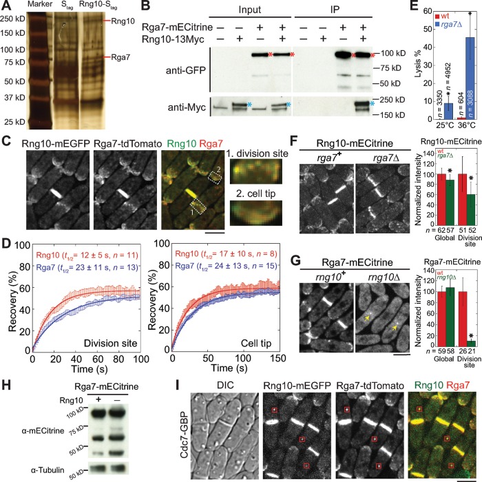
Rng10 physically interacts with the Rho-GAP Rga7 and is important for Rga7 localization. (A) Silver staining showing S. pombe proteins pulled down by Stag and Rng10-Stag. Red lines mark Rng10 and Rga7 bands. (B) Rng10 and Rga7 coimmunoprecipitate from cell extracts. Asterisks mark Rga7 (red) and Rng10 (blue). (C) Deconvoluted images showing colocalization of Rng10 and Rga7. Right, magnified regions at the division site (1) and the cell tip (2). (D) FRAP analyses of Rng10-mECitrine and Rga7-mECitrine at the division site (left) and the cell tip (right). Cells were bleached at time 0. Mean ± 1 SEM. (E) Quantification of cell lysis in wt and rga7Δ cells at 25°C and 4 h at 36°C. *p < 0.05 compared with wt. (F, G) Localization (left) and protein level (right) of Rng10 (F) and Rga7 (G). Arrows in G mark the Rga7 dot remaining at the division site. *p < 0.0001 compared with wt. (H) Western blotting showing Rga7 protein levels in cells extracts from wt (+) and rng10Δ (–) cells. Tubulin was used as a loading control. (I) Rng10 mislocalizes Rga7 to SPBs (boxes) using Cdc7-GBP. Bars, 5 μm.

