
FIGURE 8:
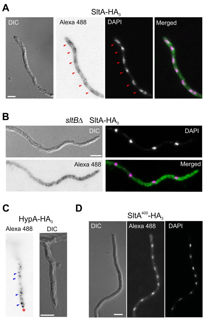
Intracellular localization of SltA. HA3 fusions were detected using a secondary antibody labeled with Alexa Fluor 488 (Alexa 488). (A) Immunofluorescence detection of SltA-HA3 expressed by strain MAD3652. (B) Detection of SltA400-HA3 expressed by null sltB strain MAD3693 under the regulation of the thiamine-repressible promoter in the absence of thiamine. (C) Detection of HypA-HA3 fusion as a quality control for immunodetection procedure. HypA-HA3 (MAD4784) displayed a similar localization to HypA-GFP, as described in Pinar et al. (2015). The red asterisk indicates the Spitzenkörper, and blue arrowheads indicate the intracellular vesicles, probably the Golgi. (D) Detection of SltA400-HA3 expressed by strain MAD4661 under the regulation of the thiamine-repressible promoter in the absence of thiamine. Bar, 5 μm.