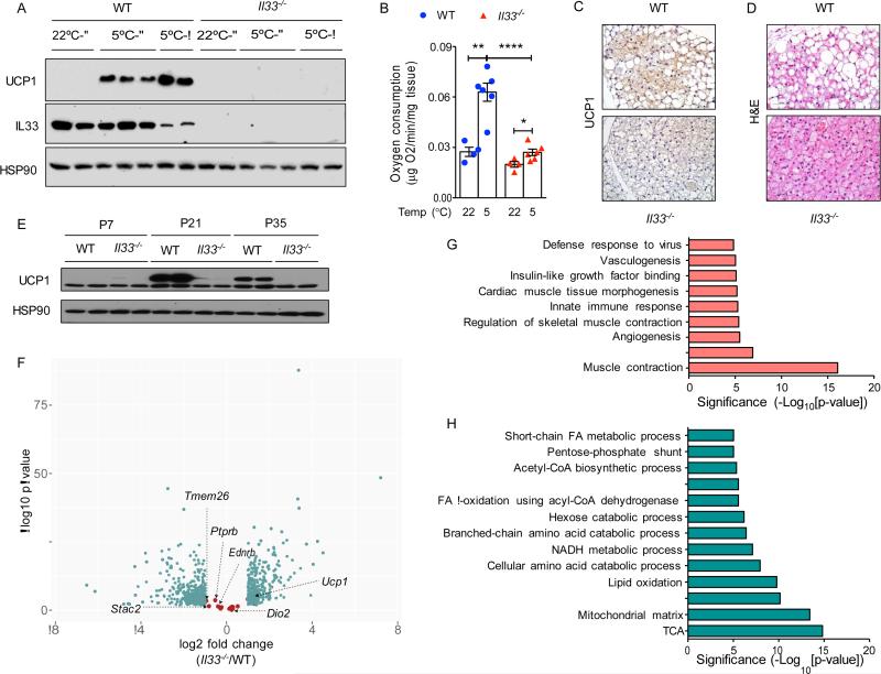
Figure 1. IL-33 is required for uncoupled respiration in beige adipocytes.
(A) Immunoblotting for UCP1 in iWAT of WT and Il33−/− male and female mice housed at 22°C or 5°C for 48 hours (n=2-3 per temperature and gender). (B) Ex vivo oxygen consumption rate in iWAT of WT and Il33−/− mice housed at 22°C or 5°C for 48 hours (n=4-6 per genotype and temperature). (C, D) Representative sections of iWAT from WT and Il33−/− mice housed at 5°C for 48 hours were stained for UCP1 (C) or with hematoxylin and eosin, 400x magnification. (E) Immunoblotting for UCP1 in iWAT of WT and Il33−/− mice housed at 22°C (n=2 per gender and age). (F) RNA-seq analysis of iWAT at P24 from WT and Il33−/− mice housed at 22°C. Volcano plot displaying fold change (log2) of mRNA expression is shown. Beige fat markers are highlighted in salmon, whereas differentially expressed mRNAs (FDR<5% and >2-fold expression) are shown in teal (n=3-4 per genotype). (G, H) Pathway analysis of a subset of mRNAs suppressed (G) or induced (H) >2-fold in P24 iWAT of WT and Il33−/− mice housed at 22°C (n=3-4 per genotype). Data are represented as mean ± SEM. See also Figure S1 and Tables S1-S3.

