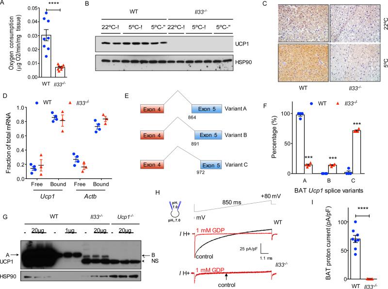
Figure 3. IL-33 is required for splicing of Ucp1 mRNA and expression of functional UCP1.
(A) Oxygen consumption rate in BAT of WT and Il33−/− mice housed at 22°C (n=8 per genotype). (B) Immunoblotting for UCP1 protein in BAT of WT and Il33−/− male and female mice housed at 22°C or 5°C (n=2-3 per temperature and gender). (C) Immunostaining for UCP1 in representative sections of BAT from WT and Il33−/− mice housed at 22°C or 5°C. (D) Polysome analysis. (E) Schematic diagram showing alternative splice variants generated from activation of cryptic splice acceptor sites in exon 5 of Ucp1. Variant A utilizes the normal splice junctions between exons 4 and 5, whereas variants B and C use cryptic splice sites in exon 5. (F) Quantification of Ucp1 splice variants present in BAT of P0.5 WT and Il33−/− mice. Analysis of Ucp1 mRNA splicing was performed using the RNA-seq datasets. (G) Immunoblotting for UCP1 in BAT of WT, Il33−/− and Ucp1−/− 0.5 day-old mice (n=3 per genotype); NS-non-specific, A-variant A, B-variant B. (H) Representative currents recorded in whole-mitoplasts prepared from WT and Il33−/− BAT. Whole-mitoplast UCP1 current before (black) and after (red) addition of 1 mM GDP to the bath solution. The voltage protocol is indicated at the top. The pipette-mitoplast diagram indicates the recording conditions. (I) Quantification of UCP1 current in whole-mitoplasts prepared from WT and Il33−/− BAT (n=7-8 per genotype). See also Figures S2 and S3.

