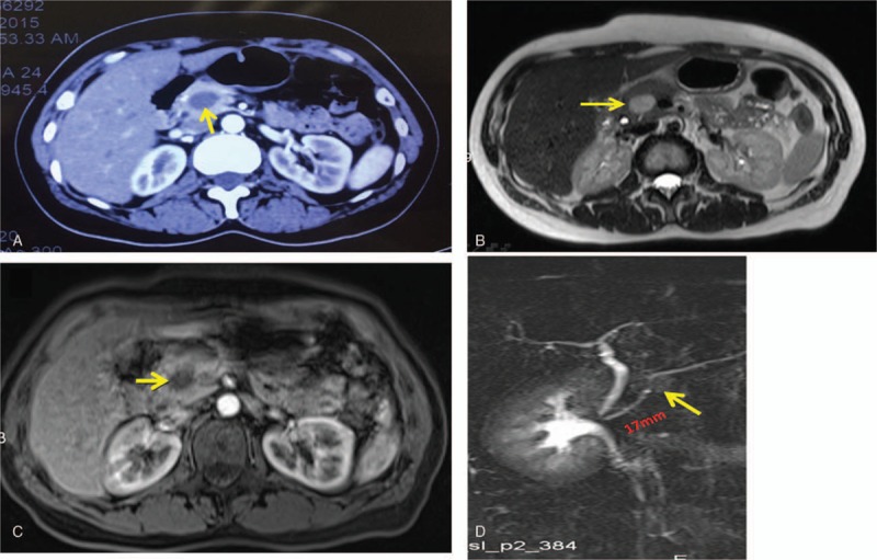
Figure 1.

CT, MRI, and MRCP scans. (A) Contrast-enhanced CT and (B, C) contrast-enhanced MRI showed a mixed density tumor in the head of the pancreas (yellow arrow). (D) MRCP showed the main pancreatic duct without dilation, approximately 1.7 cm between the mass (yellow arrow) and Vater's ampulla. CT = computed tomography, MRI = magnetic resonance imaging; MRCP = magnetic resonance cholangiopancreatography.
