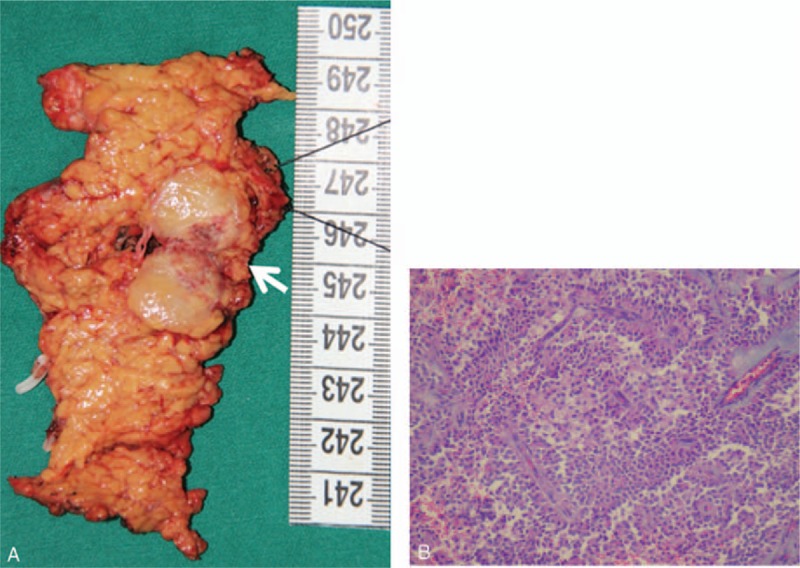
Figure 2.

(A) Gross appearance of the SPN. A well-capsulated mass, 2.2 × 1.7 cm in size, was located in the pancreatic neck (white arrowhead). (B) Histopathology of the SPN (H&E × 100). The tumor showed papillary structures with cyst-like spaces. H&E = hematoxylin & eosin, SPN = solid pseudopapillary neoplasm.
