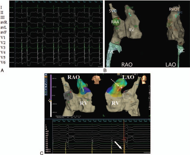
Figure 1.

(A) Surface electrocardiograms show frequent premature ventricular complexes before radiofrequency catheter ablation. (B) Path and geometry-relevant structure during catheter insertion. The name of surface leads and AB electrodes were shown in the left panel of the figure. (C) Electrophysiology study was performed without X-ray exposure guided by the Ensite NavX system. Upper panel: The yellow dot denotes the His bundle; the lowest green dot marks the tip of AB placed at target site in the septum of the right ventricular outflow tract; the red dot denotes 2 sites where ablation was attempted for 10 seconds but failed (LAO, left anterior oblique view, RAO = right anterior oblique view). Lower panel: The local ventricular activation at the tip of AB is 33 milliseconds earlier than that at surface electrocardiography recordings; radiofrequency ablation at the upper green dot abolishes the ventricular premature beat and ventricular tachycardia; from the top down are the unipolar recordings from the ablation catheter (AB), surface lead I, II, III, avR, avL, avF, and V1 to V6, and the bipolar recordings from distal pair of the AB.
