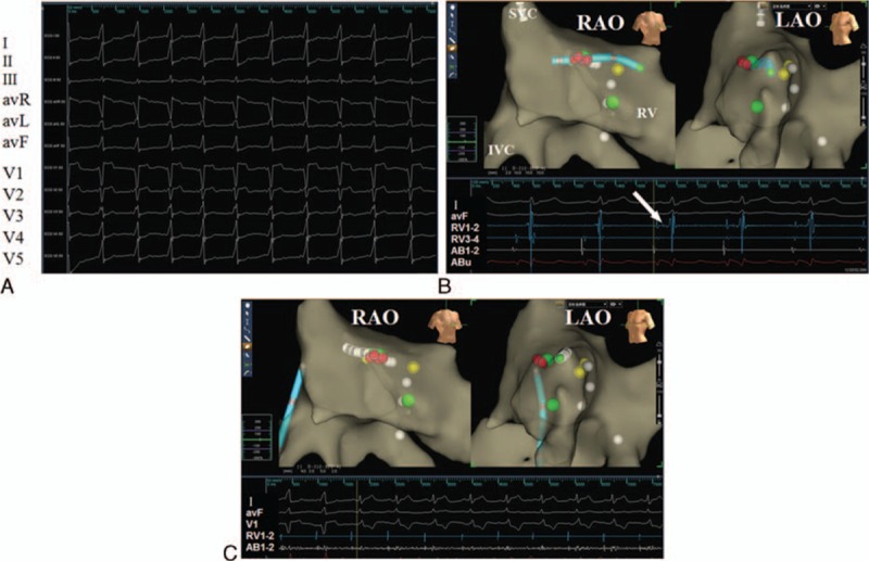
Figure 2.

(A) Surface electrocardiograms show an evident delta wave due to an accessory pathway before radiofrequency catheter ablation. (B) Electrophysiology study was performed without X-ray exposure guided by the Ensite NavX system. Upper panel: The yellow dot denotes the His bundle; the green dot denotes the suspected location of 2 accessory pathways; the tip of tetrapolar catheter was placed near the His bundle to verify the right anatomic location after the tentative ablation and before the final ablation delivery (LAO, left anterior oblique view, RAO = right anterior oblique view). Lower panel: Surface electrocardiography recordings showed the occurrence of supraventricular tachycardia (SVT); the white arrow indicates the potential of the His bundle recorded by ablation catheter (AB). (C) Radiofrequency ablation at the upper green dot abolishes the accessory pathway. The name of surface leads, RV, and AB electrodes are shown in the left panel of the figure.
