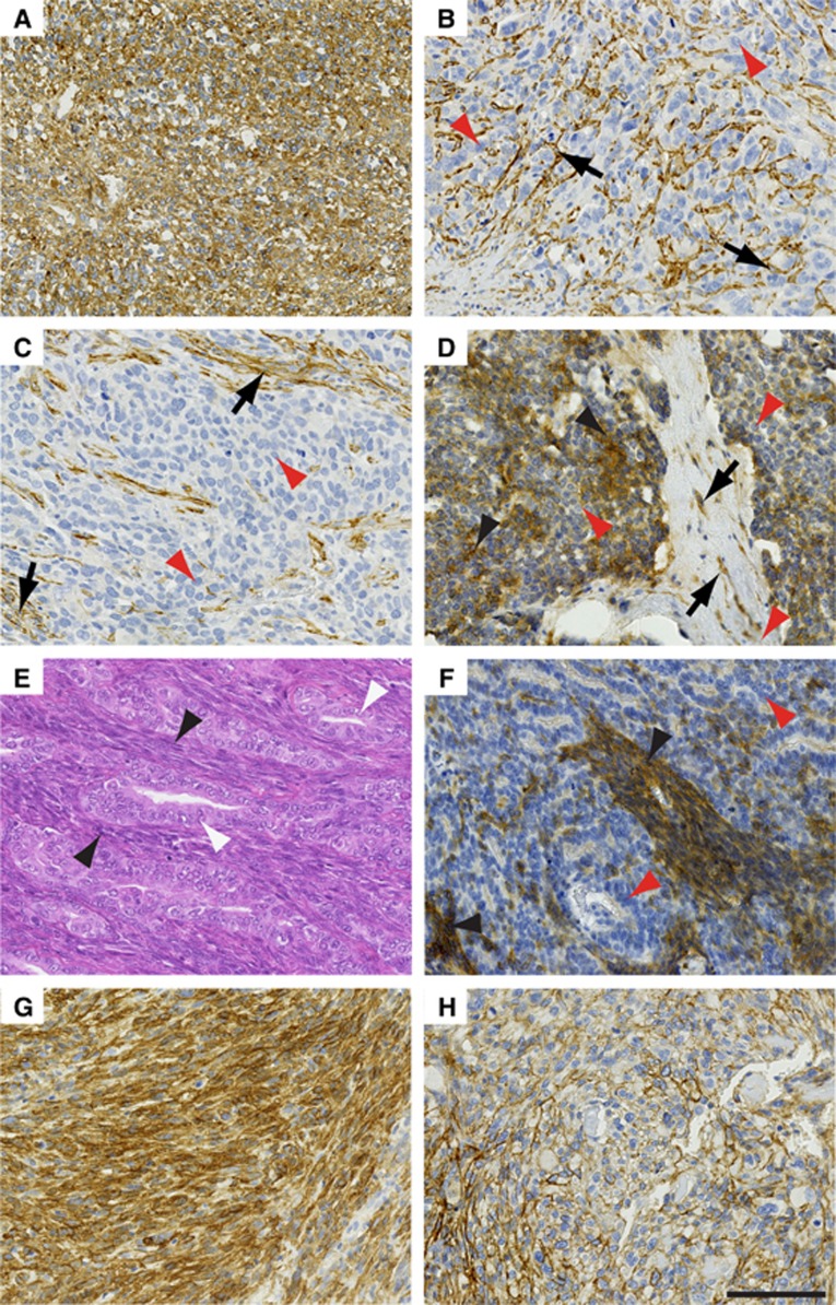
Figure 3.
Endosialin in expression in non-UPS sarcomas. (A, C) Embryonal rhabdomyosarcoma. Panel A, Example 1 showing strong uniform expression of endosialin throughout the ovoid and spindle tumour cell populations. Panel B, Example 2 where the spindle and ovoid cells of the neoplasm show no expression of endosialin (red arrowheads), but strong endosialin expression on the pericytes of the tumour vasculature (black arrows). Panel C, Example 3–as in example 2 except additional expression of endosialin on stromal fibroblasts (black arrows). (D) Alveolar rhabdomyosarcoma showing focal expression of endosialin (black arrowheads) throughout the ovoid and spindle cell population, interspersed by endosialin-negative tumour cells (red arrowheads). Characteristic fibrous septa containing endosialin-positive fibroblasts (black arrows) divide the nests of round cells. (E, F) Biphasic synovial sarcoma. Panel E, haematoxylin and eosin-stained section showing spindle cell (black arrowheads) and glandular (white arrowheads) tumour cell components. Panel F illustrates an example with strong endosialin expression within the spindle cell component (black arrowheads), whereas the glandular component, composed of more rounded tumour cells, is largely endosialin-negative (red arrowheads). (G, H) Leiomyosarcoma. Panel G illustrates a tumour with high-level endosialin expression throughout. Panel H illustrates a tumour with focal endosialin expression. Scale bar, 100 μm. See Supplementary Figure S3 for higher power images.

