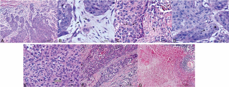
FIGURE 1.

Microscopic findings of lung squamous carcinoma (hematoxylin and eosin stain). Tumor budding is defined as a cluster of tumor cells composed of <5 tumor cells at the invasive margin of the tumor. Single cell invasion showed by arrows. Large cell invasion (arrows) was classified using the cut point of 4 lymphocytes (arrows) nearby in diameter. Mitosis was showed by arrow. Cytologic atypia was evaluated according to the size and shape of tumor cells. The severe degree was defined that the largest cell was twice larger than the smallest one. Fibrosis was evaluated using the cut point of 50%. Necrosis was evaluated using the cut point of 10%.
