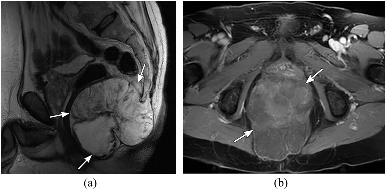
Figure 11.
A 55-year-old female with sacral chordoma. (a) Sagittal T2 weighted and (b) axial fat-saturated post-contrast T1 weighted images through the pelvis showing a large lobulated mass in relation to the coccyx (arrows) with heterogeneous high signal on T2 weighted and heterogeneous post-contrast enhancement.

