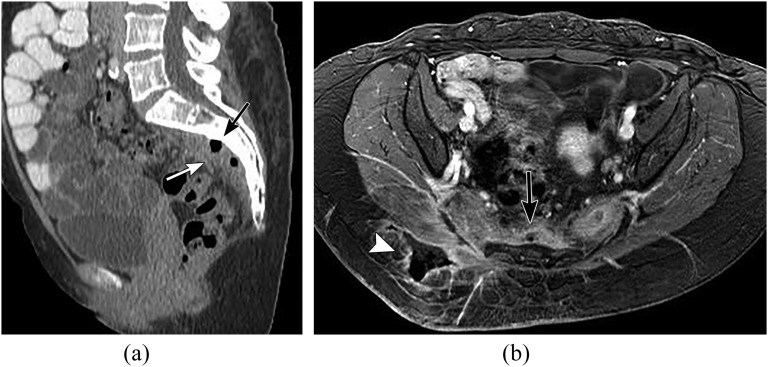
Figure 14.
A 51-year-old male with sacral osteomyelitis and presacral abscess. (a) Sagittal reformatted CT, (b) axial fat-saturated post-contrast T1 weighted image through the pelvis showing sclerosis of the sacrum (black arrow, a) with presacral soft tissue and gas (white arrow, a). MR showing phlegmonous changes in the presacral region (black arrow, b) with a fistulous track extending into the right gluteal region (white arrowhead, b).

