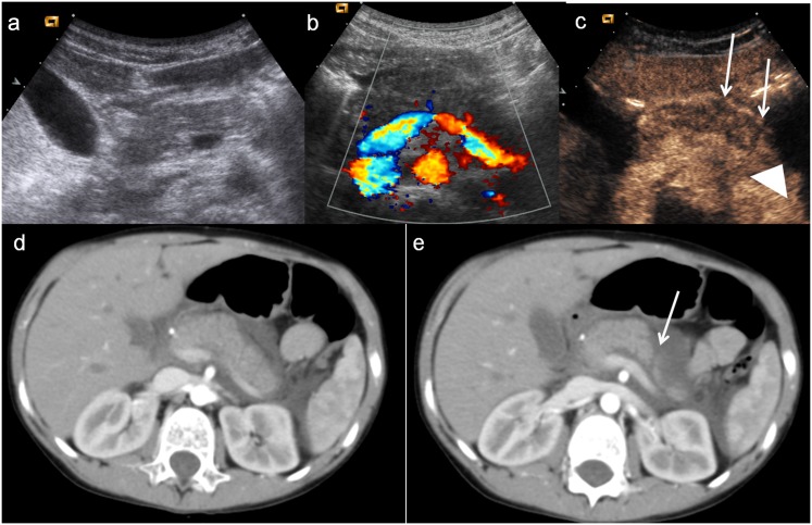
Figure 12.
Child, pancreatic injury. (a–b) Baseline ultrasonography with CD imaging does not show any pancreatic lesion. c) Contrast-enhanced ultrasound shows swelling of pancreatic body and allows to suspect subtle lesions of the pancreatic body and tail (white arrows), associated with pre-pancreatic fluid collection (arrowhead). (c–d) Axial CT scans confirm the pancreatic tail lesion (white arrow) with pre-pancreatic fluid collection.

