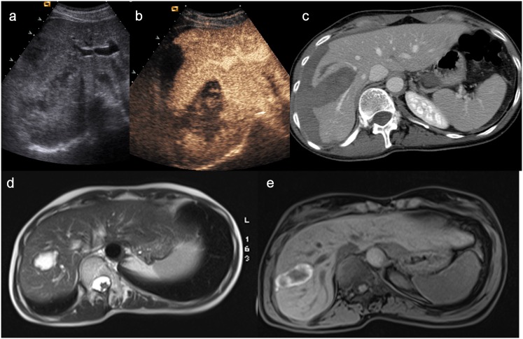
Figure 15.
(a) Baseline ultrasonography shows an inhomogeneous hypoechoic–hyperechoic parenchymal lesion of the liver and a subcapsular haematoma; (b) contrast-enhanced ultrasound (CEUS) allows to depict the hepatic laceration and the parenchymal and subcapsular haematoma well; (c) axial CT scan confirms the CEUS findings; (d–e) MRI, performed 4 months later, demonstrates the reduction in size of the lesion and the disappearance of subcapsular haematoma well.

