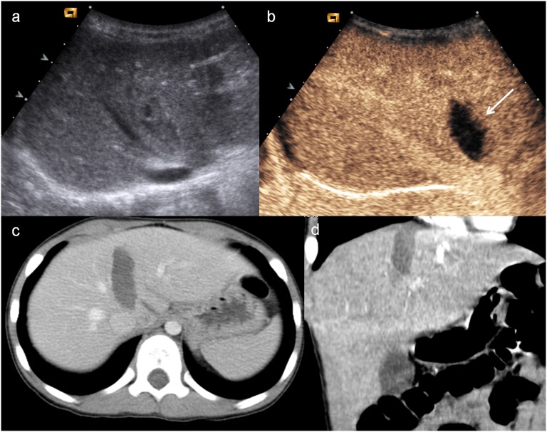
Figure 2.
Child, hepatic injury. (a) Baseline ultrasonography shows a mild hyperechoic area in the liver parenchyma; (b) contrast-enhanced ultrasound (CEUS) demonstrates a well-defined hypoechoic lesion (white arrow); (c) axial scan and (d) coronal CT reconstruction confirm the hepatic lesion, corresponding in size and shape to that observed at CEUS.

