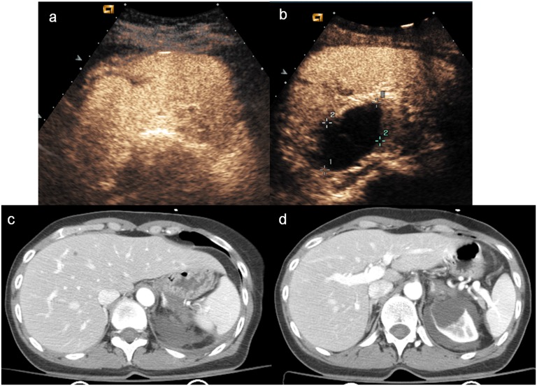
Figure 9.
Females, splenic and renal injury. (a–b) Contrast-enhanced ultrasound shows a subtle lesion of splenic parenchyma, involving the capsule, which was not evident on baseline ultrasonography. A large cyst of renal upper pole is also depicted; (c–d) axial CT scan confirms the little lesion of the spleen and a subcapsular renal haematoma, due to the rupture of the upper pole cyst.

