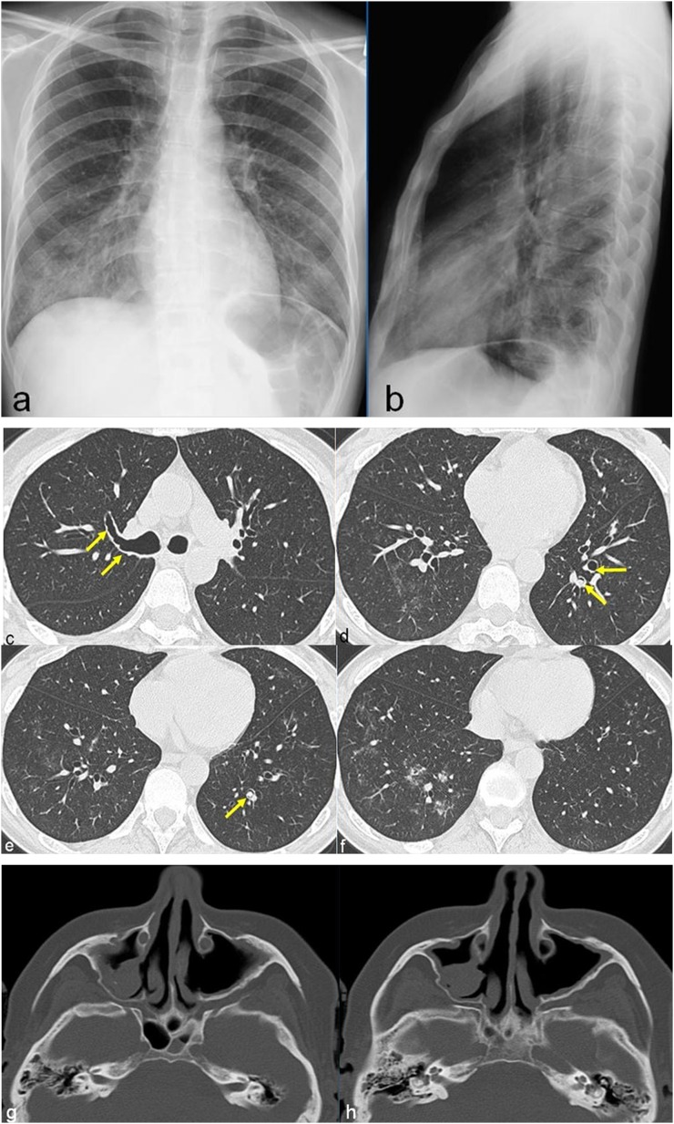
Figure 8.
A 50-year-old female with allergic asthma, rhinitis and peripheral neuropathy. Chest radiographs show hazy areas of increased opacity prominent in the middle and right lower lobes (a, b). CT images, obtained after 10 days from chest radiographs, show diffuse and irregular bronchial wall thickening (arrows in c) associated with the presence of nodules with endo- (arrows in d, arrow in e) and peribronchial (f) distribution. CT of the facial bones shows bilateral meatal antrostomy of maxillary sinuses with signs of chronic sinusopathy (g, h).

