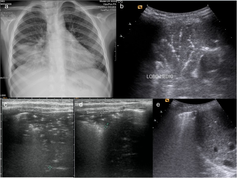
Figure 1.
A 6-year-old boy with cough and fever. (a) Chest X-ray (CXR) performed at admission in anterior–posterior projection shows an extensive medial lobe consolidation; (b) lung ultrasound (LUS) performed simultaneously with CXR shows a typical pneumonic consolidation with “arborescent bronchogram”; (c) in association, there was a wider consolidation with “parallel” bronchogram suggesting a prevalent atelectasis component, measures were taken to follow up it up; (d) follow-up at 7 days. LUS shows a significative reduction in size of lung consolidation, with persistent “parallel” bronchogram suggesting residual minimal atelectasis component; (e) follow-up at 14 days. LUS shows complete resolution of lung consolidation, with residual pleural line thickening.

