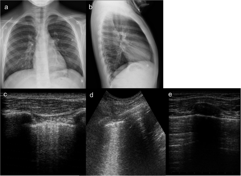
Figure 6.
A 11-year-old boy presenting with mild dyspnoea, cough and fever. (a, b) Chest X-ray does not show any consolidation or interstitial disease. (c, d) Lung ultrasound performed simultaneously on lung bases depicts well multiple “B-lines”, ring-down, vertical artefacts, expression of interstitial involvement, so much different from the normal pattern of “A-lines”. (e) Artefacts parallel to pleural lines, which are present at upper lobes.

