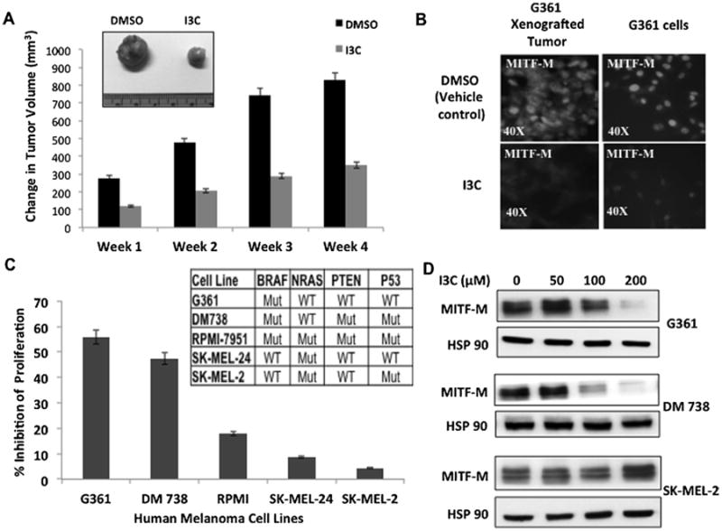
Figure 1.

Effects of I3C on in vivo growth of melanoma tumor xenografts, production of MITF-M, and melanoma cell proliferation. (A) Athymic mice with G-361 cell-derived tumor xenografts were injected subcutaneously with either I3C or with DMSO vehicle control, and resulting tumor volumes were calculated as described in the Supporting Information. The micrograph insert shows tumors harvested at the end of week 4. (B) At terminal sacrifice, tumor sections were analyzed for MITF-M expression by immunofluorescence using primary antibodies to MITF-M (left panel). Cultured G361cells treated with 200 μM I3C for 72 h were similarly probed for MITF-M levels (right panel). (C) Human melanoma cell lines with distinct genotypes were treated with or without 200 μM I3C for 48 h and the effects on cell proliferation measured using a CCK-8 assay relative to the vehicle control. (D) The levels of MITF-M protein were determined in melanoma cells treated with the indicated concentrations of I3C for 48 h by western blots.
