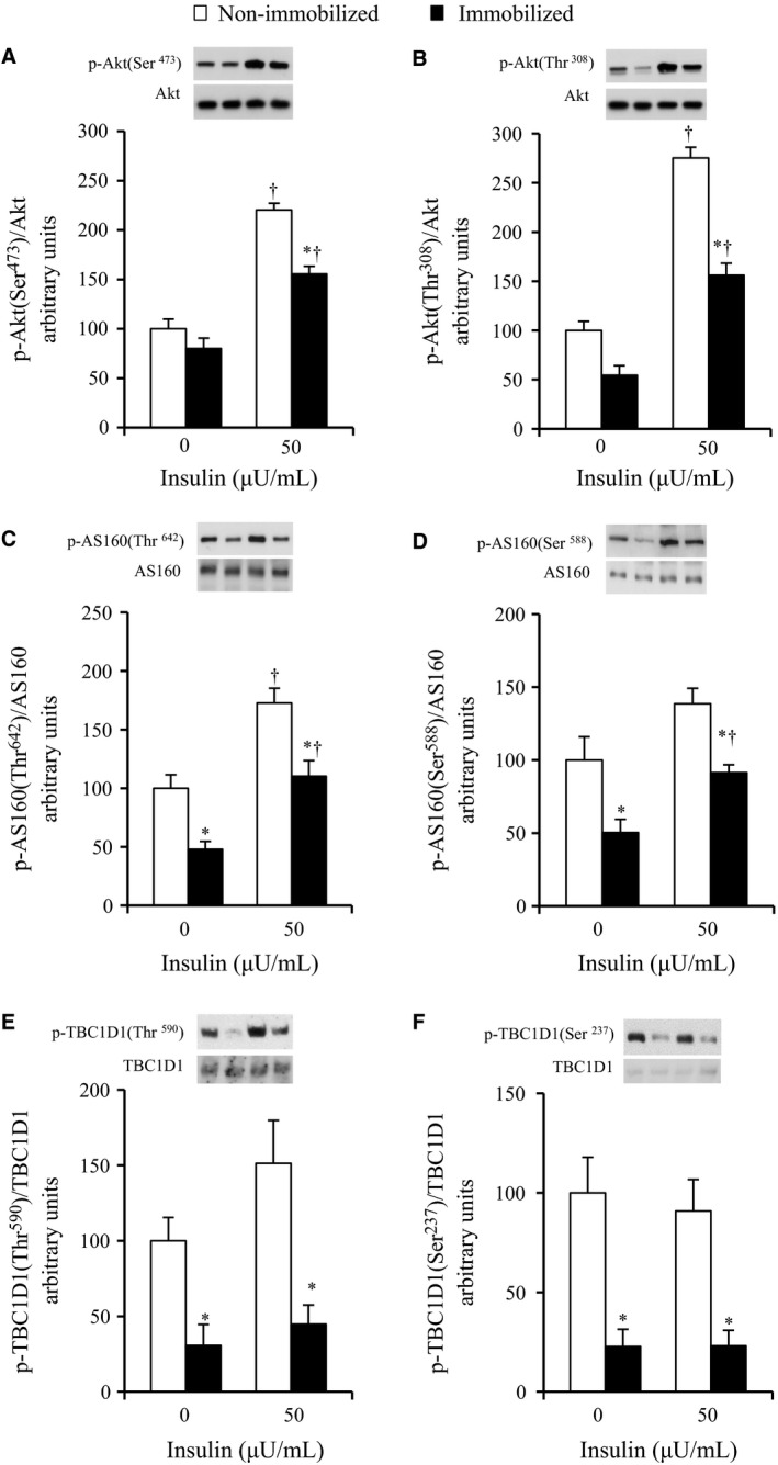
Figure 2.

Phosphorylations of Akt, AS160, and TBC1D1 in contralateral non‐immobilized and immobilized limbs at the end of 6‐h hindlimb immobilization. Muscles were dissected out at the end of 6‐h unilateral hindlimb immobilization. All muscles were incubated in glucose‐free medium in the absence or presence (50 μU/mL) of insulin for 20 min and then frozen. Muscle lysates were separated with SDS‐PAGE and blots were analyzed for phosphorylated Akt Ser473 (A), phosphorylated Akt Thr308 (B), phosphorylated AS160 Thr647 (C), phosphorylated AS160 Ser588 (D), phosphorylated TBC1D1 Thr590 (E), and phosphorylated TBC1D1 Ser237 (F). Blots were then stripped and analyzed for total abundance of each protein. (A–C, F) Values are means ± SE (n = 7–9). (D–E) Values are means ± SE (n = 6–7). *P < 0.05 versus the contralateral non‐immobilized limbs with the same insulin concentration. † P < 0.05 versus 0 μU/mL insulin.
