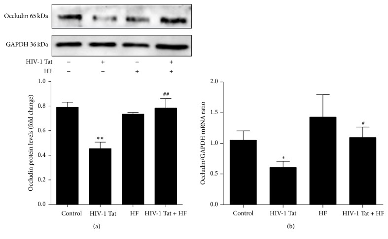
Figure 2.
Role of RhoA/ROCK signaling in HIV-1 Tat-induced changes in occludin. hCMEC/D3 cells were pretreated with HF (30 μmol/L) 2 h prior to the addition of occludin. HIV-1 Tat treatment was continued for 24 h for western blotting (a) and for 12 h for RT-PCR (b). HIV-1 Tat exposure was associated with decreased protein and mRNA levels of occludin in hCMEC/D3 cells. With coexposure to HF and HIV-1 Tat, occludin protein and mRNA levels were significantly increased when compared with exposure to HIV-1 Tat only. Data are expressed as means ± standard error of the mean (n = 3, values determined by the ratio to GAPDH, for (a), n = 5 for (b)). ∗ p < 0.05 and ∗∗ p < 0.01 versus control; # p < 0.05 and ## p < 0.01 versus HIV-1 Tat.

