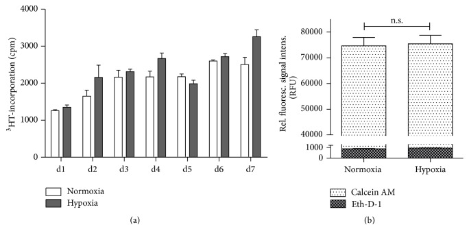
Figure 1.
HeLa-hTDO cells survive incubation under hypoxic conditions. (a) Proliferation assay: 1 × 10³ HeLa-hTDO cells were seeded per cm2 in 25 cm2 cell culture flask (Corning, NY, USA), 3H-thymidine was added at day 0, and the cells were incubated under normoxia (20% O2) or hypoxia (1% O2) for 1–7 days. The incorporation of 3H-thymidine was detected using liquid scintillation spectrometry (1205 Betaplate, PerkinElmer, Rodgau Jügesheim, Germany). (b) Fluorescence-based cell viability/cytotoxicity assays: 3 × 10⁴ HeLa-hTDO cells per well were incubated for 24 h under normoxia (20% O2) or hypoxia (1% O2). Then the cells were stained with Calcein AM (white bars) or Ethidium-Homodimer-1 (Eth-D-1) (plaid bars) indicating living or dead cells, respectively. After incubation the fluorescence was detected using a fluorescein optical filter (excitation 485 ± 10 nm; emission 530 ± 12 nm) for Calcein or using a rhodamine optical filter (excitation 530 ± 12 nm; emission 645 ± 12 nm) to detect Eth-D-1. For both assays a two-tailed unpaired t-test was used to compare the groups (n.s. = not significant), n = 3 independent experiments with three replicates each. The bars indicate the mean value ± SEM.

