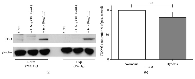
Figure 3.
Unaltered hTDO expression in HeLa-hTDO cells under hypoxic conditions. (a) Exemplary Western Blot protein analysis of hTDO and β-actin protein expression in HeLa-hTDO cells after 72 h of incubation under normoxia (20% O2) or hypoxia (1% O2). (b) Densitometric evaluation of Western Blot protein analyses: ratio of relative hTDO protein expression to β-actin protein expression as % of positive control ± SEM, n = 8 independent experiments. Comparison of hTDO/β-actin protein ratio under normoxia or hypoxia via a two-tailed, paired t-test; n.s. = not significant.

