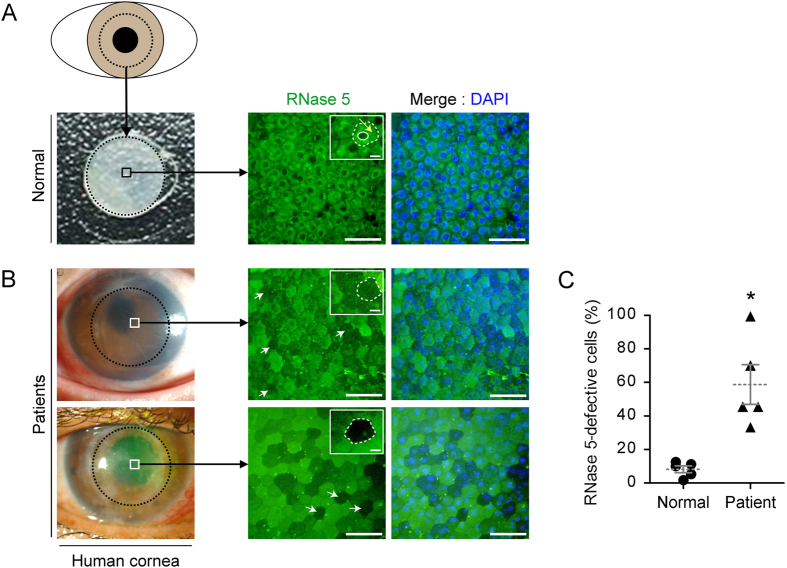
Figure 1. Ex vivo expression patterns of ribonuclease (RNase) 5 protein in normal and decompensated human corneal endothelial tissues.
(A,B) Representative images showing flat-mounted immunofluorescence (IF) staining of RNase 5 protein in normal (A) and decompensated human corneal endothelial tissues (B). Central circular corneal tissues with a diameter of approximately 8 mm (dotted black circles) were punched and excised from donor corneas. Thereafter, RNase 5 protein-expressing corneal endothelial cells (CECs) in the central area of corneas (squares in left column) were microscopically evaluated by IF. Protein expression of RNase 5 was clear in CECs of normal tissue, but defective in CECs of decompensated tissues (white arrows, shown representatively). In the magnified views (small white rectangles in middle column), normal CECs exhibited copious expression of RNase 5 protein in the cytoplasm (yellow arrow, A) between the CEC contour (dotted white circle) and nucleus (white solid circle). On the other hand, the expression of RNase 5 protein was defective by IF stain in CECs in decompensated human corneal endothelial tissues (dotted white circles, B). (B) Photos in the left column are snapshots of a cornea taken prior to surgical excision for corneal transplantation. Scale bar: 50 μm except for those in small white rectangles, which are 10 μm. (C) The proportion of CECs that exhibited defective expression of RNase 5 protein according to IF analysis was significantly higher in corneal tissues from patients with decompensated corneal endothelium. *p = 0.012, vs. normal (t-test). n = 5 corneas in each group. Values represent the mean ± s.e.m.

