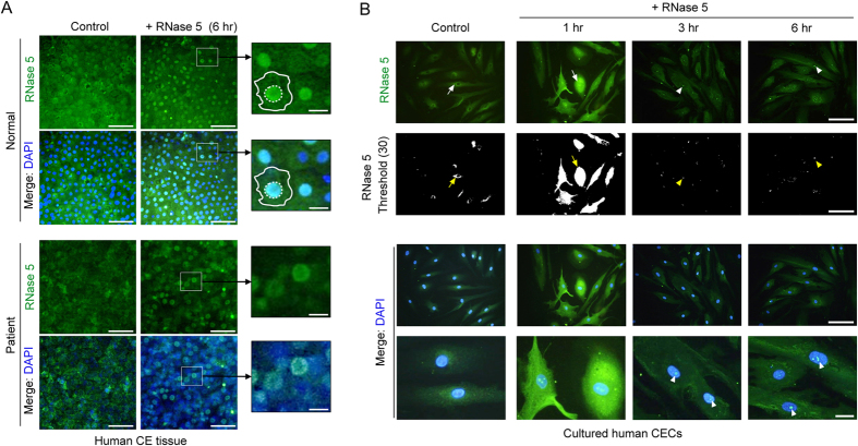
Figure 4. Ribonuclease (RNase) 5 treatment-induced nuclear localization of RNase 5 in human corneal endothelium tissues and cultured human corneal endothelial cells (CECs).
(A) Representative images showing flat-mounted immunofluorescence stain of RNase 5 in normal and decompensated human corneal endothelial tissue with or without RNase 5 treatment. RNase 5 was predominantly expressed in the nucleus after 6-hour treatment with exogenous human RNase 5 (5 μg/mL) in normal and decompensated corneal endothelium (right column). In magnified images of small white rectangles, the contour of CECs (white border lines) and the nuclear expression of RNase 5 after RNase 5 treatment (white dotted circles) are outlined. Scale bar: 50 μm (white) and 10 μm (yellow). n = 3 independent experiments. (B) RNase 5 localization in cultured human CECs with or without RNase 5 treatment. RNase 5 expression was detected at the cytoplasmic perinuclear area in untreated CECs (arrows in the control column). After RNase 5 treatment, intracellular RNase 5 expression became very prominent early (arrows in 1 hr column), and thereafter localized specifically to the nuclei (arrow heads in 3 hr and 6 hr columns). This observation was more apparent with thresholding (upper second row, lightness 30). Scale bar: 100 μm (three upper rows), 20 μm (lowest row). n = 3 independent experiments.

