
Figure 4.
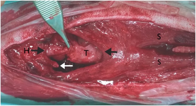
Intraoperative pictures; ventral approach to the larynx: the head is on the left side of the picture. Reattachment of the ventral epiglottic mucosa, fracture of the right thyrohyoid bone (white arrow), basihyoid bone (H), thyroid cartilage (T), and sternohyoid muscle (S), the black arrows show the ends of the muscle tissue that were sutured back together.