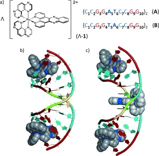
Figure 1.

a) Structures of Λ‐[Ru(TAP)2(dppz)]2+ and ODNs used in this study. b,c) Crystal structures of Λ‐[Ru(phen)2(dppz)]2+ bound to b) ODN A and c) ODN B from Niyazi et al.,5b showing binding at central T5A6:T5A6 step in {CCGGTACCGG}2 but not {CCGGATCCGG}2. Intercalated complexes only are shown. Color code : Guanine, red; cytosine, cyan; adenine, green; thymine, yellow; nitrogen, blue.
