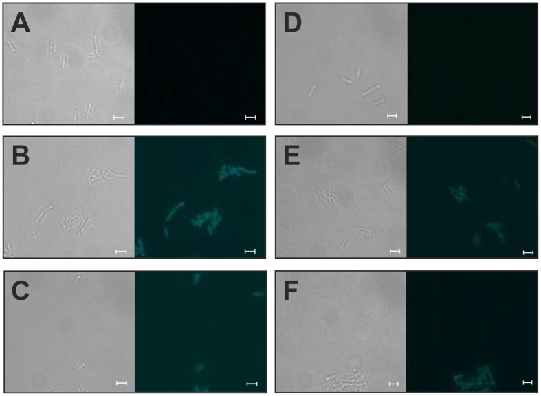
Figure 1.
Fluorescence microscopic analyses of C. ljungdahlii wild type (A), C. ljungdahlii (pGlow-CKXNBs2) (B), C. ljungdahlii (pGlow-CKXNPp1) (C), C. acetobutylicum wild type (D), C. acetobutylicum (pGlow-CKXNBs2) (E) and C. acetobutylicum (pGlow-CKXNPp1) (F). The left panels represent light microscopic images and the right panels show fluorescence microscopic images collected with a Leica DFC 365 FX fluorescence microscope equipped with a fluorescence cube 405 at excitation wavelength from 375–435 nm and emission wavelength of 445–495 nm. Scale bars, 3.23 μm.

