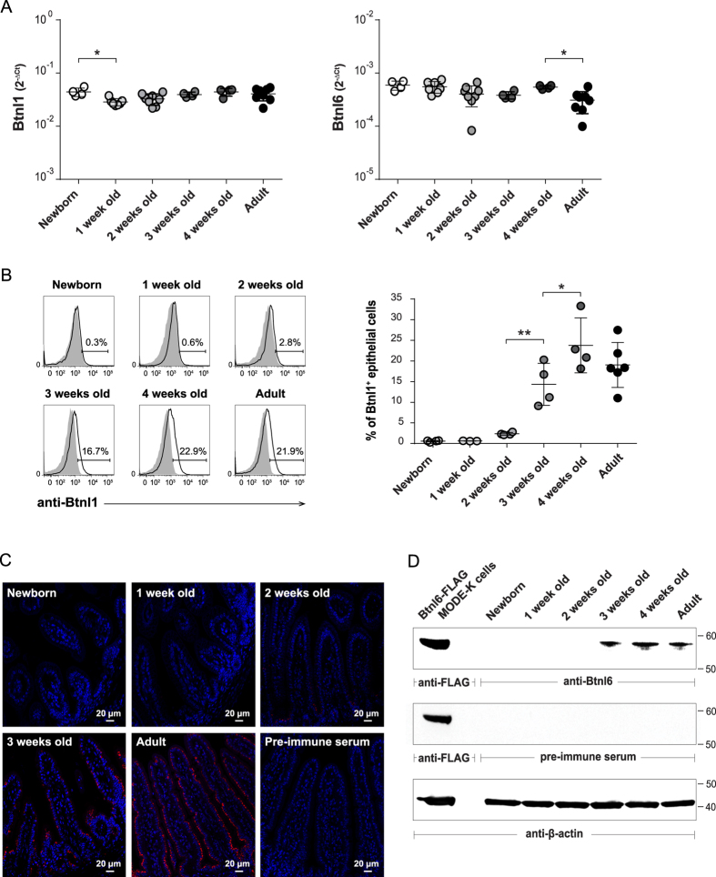
Figure 1. Btnl1 and Btnl6 expression in murine intestine during ontogeny.
Expression of Btnl1 and Btnl6 genes (A), and Btnl1 (B,C) and Btnl6 proteins (D) was examined in small intestinal tissue of newborn (day 0), 1, 2, 3, and 4-week-old and adult C57BL/6 mice. (A) Btnl1 and Btnl6 gene expression was assessed by qPCR, run in duplicates, and normalized against β-actin. (B) Murine intestinal epithelial cells, gated on CD45− epithelial cells and 7AAD negative cells to exclude non-viable cells, were stained with anti-Btnl1 rabbit polyclonal antiserum (solid black line) or pre-immune serum (shaded histogram) which served as a negative control. Representative histogram for each time-point is shown. 4–8 mice/time-point were analyzed and One-Way ANOVA followed by Holm-Sidak´s multiple comparisons test was used for statistical analysis (*P ≤ 0.05, **P ≤ 0.01, ***P ≤ 0.001 and ****P ≤ 0.0001) (A,B). (C) Small intestinal sections were immunostained with anti-Btnl1 rabbit polyclonal antiserum (red) and counterstained with DAPI (blue) to visualize nuclei. No staining was detected using pre-immune serum. Original magnification 20x. Four mice were stained for each time-point and representative stainings are shown. (D) Isolated iECs from small intestinal tissue of C57BL/6 mice (20 μg) were analyzed for Btnl6 protein expression. Lysates from MODE-K cells transfected with FLAG-tagged Btnl6 cDNA pMX-IRES-GFP served as a positive control. The predicted protein migrating under reducing conditions at the theoretical molecular weight of ~59 kDa for FLAG-tagged Btnl6 and ~58 kDa for non-tagged Btnl6 was detected with anti-FLAG antibody or Btnl6-specific polyclonal antibody. No bands were detected on gels immunoblotted with pre-immune serum. The β-actin immunoblot acts as a loading control. Data are representative of four experiments.

