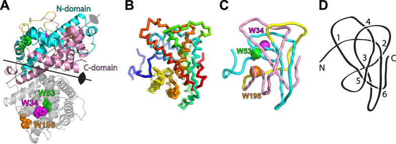
Figure 1. Structure and knot topology for DehI.
(A) Cartoon representation of the crystal structure of DehI (PDB code: 3BJX). The N- and C-terminal domains of one subunit are colored in cyan and pink, respectively, and the connecting linker in yellow. The other subunit is colored in grey with the three tryptophan residues labeled with their respective sequence numbers. The two-fold symmetry axes of the homodimer and two symmetric domains are indicated by arrows. (B) Simplified backbone representation of a DehI monomer. The ribbon diagram is color-ramped from blue to red from the N- to C-termini. (C) Reduced backbone topology of DehI generated by the pKNOT web server using the Taylor smoothing algorithm7. The positions of the three tryptophan residues are indicated. (D) The simplified 61 knot topology of DehI with the positions of the six crossings indicated.

