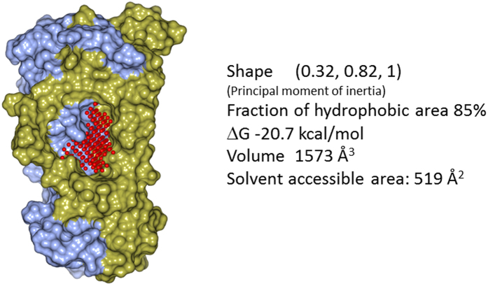
Figure 2. Characterization of the central binding pocket of the IL-17A dimer (surface presentation with the two polypeptide chains colored in ice blue and gold, respectively) probed using the VISM algorithm (red balls represent the probes used).
The high druggability of the pocket is manifested by the large hydrophobic cavity and the favorable druggability score (∆G) which assesses the optimal binding affinity of the binding site.

