
FIG 6.
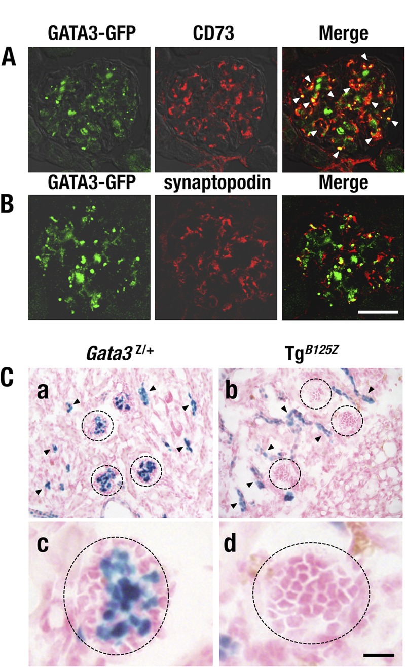
GATA3 expression in the renal glomerulus. (A) GATA3-GFP-positive cells (green) are colocalized with CD73-positive (red) mesangial cells (arrowheads in the merged image). (B) GATA3-GFP (green) and synaptopodin (red) immunoreactivities are rarely colocalized. (C) X-Gal staining of kidney from Gata3Z/+ (left) and TgB125Z (right) mice. Gata3Z/+ mice show lacZ activity-positive cells in the renal tubular cells (arrowheads) and the central region of the glomerulus (circles) (a and c). TgB125Z mice show lacZ-positive cells only in the tubules (arrowheads) (b and d). lacZ activity is largely missing from the glomeruli of TgB125Z mice. Bars, 50 μm.