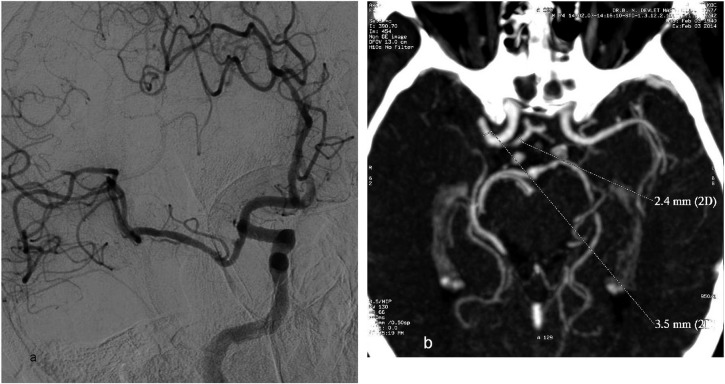
Figure 11.
Vasospasm at the recanalized middle cerebral artery (MCA) M1 segment is seen on the oblique digital subtraction angiography image (a) of a 74-year-old male patient with right MCA stroke who underwent detached stent extraction after repeated stent-assisted thrombectomy (SAT). CT angiography maximum intensity projection image (b) showing the initial diameter of the MCA M1 segment (3.5 mm) and anterior cerebral artery A1 segment (2.4 mm) before SAT, which supports the presence of vasospasm after the intervention. 2D, two dimensions.

