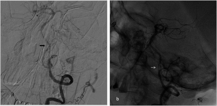
Figure 3.
Digital subtraction angiography (a) and unsubtracted (b) images showing detachment of the thrombectomy stent at the vertebrobasilar junction in a 63-year-old male with basilar dissection. (a) Arrow shows the distal marker of the detached stent. (b) At the basilar artery, there was a flap appearance and subintimal contrast filling (arrow) which was highly possible for the dissection. In this patient, during the procedure, dissection progressed downwards.

