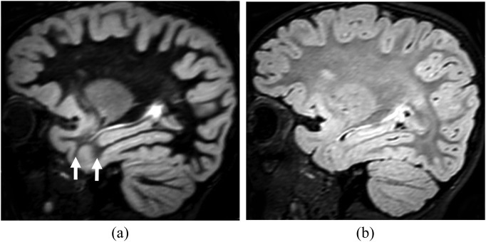
Figure 3.
Incomplete myelination in a 20-month-old child. Sagittal reformatted double inversion recovery (DIR) (a) and sagittal T2 fluid-attenuated inversion-recovery (b) images show relatively increased signal intensity in the white matter of the anterior temporal lobes when compared with the frontal white matter. This difference, due to incomplete myelination, is most conspicuous on the DIR image (arrows). Despite the apparent blurring of the grey–white matter junction, this is a normal finding when symmetric.

