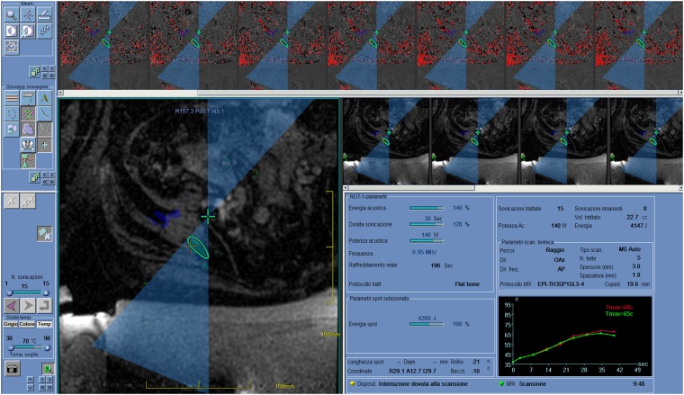
Figure 2.
Screenshot of a MRI-guided focused ultrasound surgery workstation after “sonication”, during the treatment of a painful bone metastasis from thyroid cancer in a 60-year-old female. The beam path is shown, with “near field” before the target (circle) and “far field” beyond. Typically, in bone applications, the focus (cross) is set beyond the cortical surface target to exploit the high absorption of the cortical bone. Areas/volumes covered and ablated by previous sonications are highlighted. The higher line shows thermal images during sonication. The graph at the bottom right shows the evolution of temperature during sonication, and this can be checked pixel by pixel.

