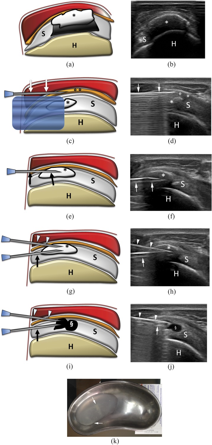
Figure 2.
Percutaneous treatment of calcific tendinopathy of the supraspinatus using double-needle technique. (a) Scheme and (b) ultrasound image of a hard calcification (asterisks) with acoustic back shadow. (c) Scheme and (d) ultrasound image of needle (arrows) insertion into the subacromial–subdeltoid bursa (circles) to inject anaesthesia. The calcification (asterisks) is visible under the reverberation artefact of the needle. (e) Scheme and (f) ultrasound image of first needle (arrows) insertion in the calcification (asterisks). Note that the bevel is open upward. (g) Scheme and (h) ultrasound image of the second needle (arrowheads) insertion in the calcification (asterisks) on the same coronal plane of the first needle (arrows). Note that the bevel of the second needle is open downward and that the back shadow of the more superficial needle partially covers the image of the deeper needle. (i) Scheme and (j) ultrasound image at the end of the procedure. The calcification is completely empty (§). (k) Saline used to wash a calcification with a double-needle procedure collected in a bowl. Note the whitish fragment of calcium dispersed in the solution (arrows). H, humerus; S, supraspinatus tendon.

