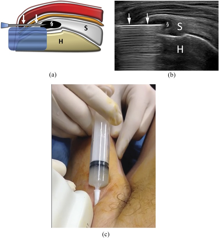
Figure 3.
Percutaneous treatment of calcific tendinopathy of the supraspinatus using single-needle technique. Bursa anaesthesia and needle insertion is similarly performed to what happens for double-needle procedure (Figures 2c–f). (a) Scheme and (b) ultrasound image at the end of the procedure performed with one needle (arrows). The calcification is completely empty (§). (c) Image taken during a single-needle procedure. The syringe is filled with whitish saline solution, containing the calcium that is being removed. Note that the luer is higher than the rest of the syringe, thus, allowing for calcium deposition on the bottom. H, humerus; S, supraspinatus tendon.

