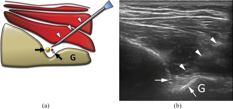
Figure 7.
(a) Scheme and (b) ultrasound image of suprascapular nerve block at the spinoglenoid notch. The needle (arrowheads) is inserted with lateral, in-plane approach to place the tip around the spinoglenoid notch (arrows), where the suprascapular neurovascular bundle runs. As the bundle is barely visible, particular caution should be taken when performing this procedure. G, glenoid.

