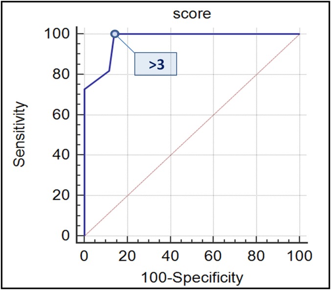
Figure 6.
Receiver operating characteristic (ROC) analysis of MRI score to discriminate MCAs from MCACs. The score was assessed for each patient by giving 1 point for each of the following high-risk MRI features: size >7 cm; thickness of septa >3 mm; thickness of the wall >3 mm; number of loculations >4; nodules; T1 hyperintensity of the cystic content; compression of adjacent organs; and metastases. Area under the curve (AUC) of the ROC curve analysis with 95% confidence limit (AUC = 0.971 and confidence interval: 0.897–0.997) revealed that the best cut-off value to discriminate MCAs from MCACs was >3 points (Youden index = 0.8605, p < 0.0001). At this cut-off value, the sensitivity, specificity, accuracy, postive-predictive value and negative-predictive value were 100%, 86%, 91%, 79% and 100%, respectively. MCAs, mucinous cystoadenomas; MCACs, mucinous borderline tumours; and cystoadenocarcinomas.

