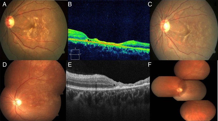
Figure 2.
At presentation pigmentary changes with fine yellowish golden dots were seen at the macula of the left eye in case 1 (A). Optical coherence tomography (OCT) of the same eye revealed significant retinal atrophy at the macula with few intraretinal cysts at the nasal margin of the atrophic area. Few hyper-reflective dots suggestive of cells were seen in the posterior vitreous (B). Within 3 days the yellowish golden deposits at the macula of the left eye started reducing, the area of retinal pigment epithelial alteration started becoming more localised with distinct demarcation (C). The left eye of case 2 at presentation showed pigmentary changes, punctate intraretinal haemorrhages at the macula with confluent patches of granular pigmentary changes spreading superotemporally (D). Corresponding OCT showed atrophy at the macula with few intraretinal cysts at the margin (E). At the 2nd week, the left eye showed healed retinitis both at the posterior pole and in the periphery (F).

