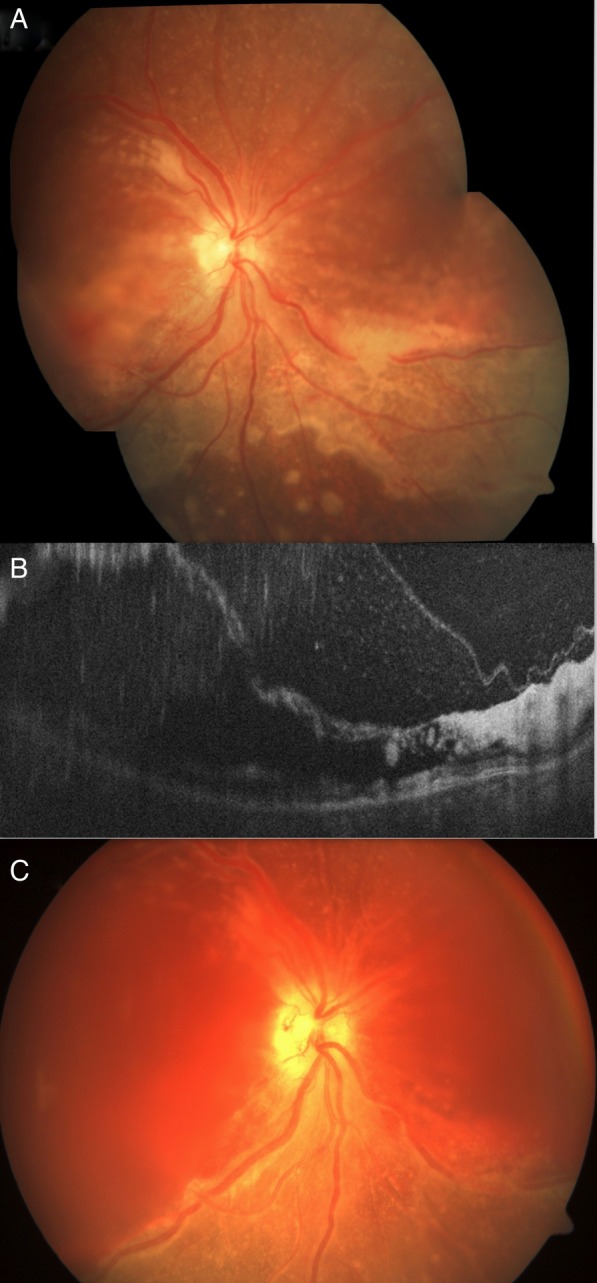
Figure 3.

The right eye of case 2 at presentation showed thin elevated retina at the posterior pole with whitish discolouration, mild intraretinal haemorrhage inferior to the macula with centrifugally spreading retinitis with an active anterior margin. Few separate multifocal round active whitish lesions were also seen beyond the central area of involvement (A). The optical coherence tomography of the right eye revealed intraretinal schisis with hyper-reflective inner retinal layers and accumulated cells between the inner retina and posterior hyaloid (B). At 4th day, case 2 presented with haemorrhage in the schitic cavity at the posterior pole with necrotic retina and multiple breaks, vitreous haemorrhage but resolving retinitis (visible inferonasal part of fundus) in the right eye (C).
