
Fig. 1.
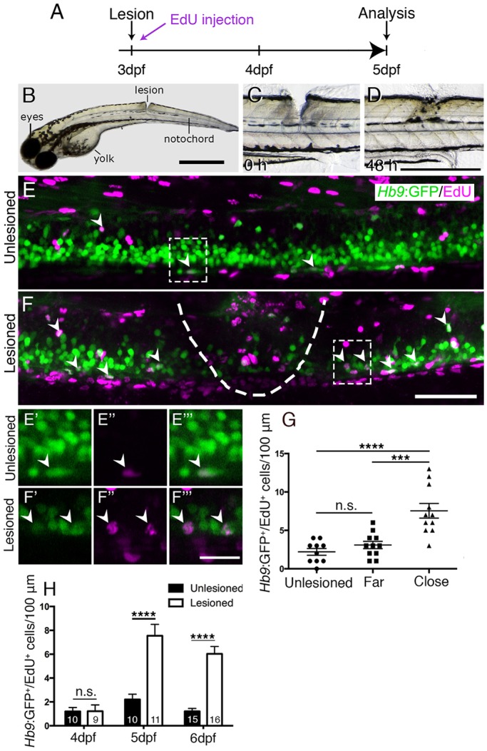
A mechanical lesion to the spinal cord heals within 48 h and leads to motor neuron regeneration close to the lesion site. (A) Time line for experiments. (B) A zebrafish larva with a lesion in the dorsal trunk area, leaving the notochord intact at 3 dpf. (C,D) The same larva imaged at 0 and 48 h post lesion shows closure of the wound. (E,F) The lesion area is outlined with double-labelled Hb9:GFP+/EdU+ neurons indicated by arrowheads. A lesion leads to increased numbers of Hb9:GFP+/EdU+ double-labelled motor neurons (compare E and F). E′ to F‴ show higher magnifications of areas boxed in E and F, respectively, in single optical sections indicating double labelling. (G) Motor neurons are regenerated close to (<50 µm rostral and caudal), but not far from (100 µm>×>50 µm rostral and caudal) the lesion site (one-way ANOVA with Bonferroni's multiple comparisons test, ***P<0.001, ****P<0.0001; unlesioned, n=10; far, n=11; close, n=11). (H) Time line of the increase in the number of EdU-labelled motor neurons (t-test, ****P<0.0001). Values are means±s.e.m. Lateral views of larvae are shown (rostral is left; dorsal is up). Scale bars: 100 µm in B; 500 µm in D for C,D; 50 µm in F for E,F; 15 µm in F‴ for E′-F‴.