
Fig. 1.
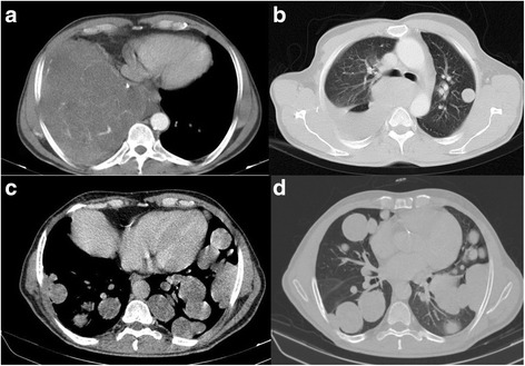
Contrast enhanced axial CT scan of the patient before a, b and after c, d surgery. There is a large solid mass in lower zone of right hemithorax with complete collapse of right lower lobe and cardiac displacement to left a. There is also an 18 × 15 mm parenchymal nodule in left upper lobe. Several other smaller nodules are found in this exam (not shown) in the rest of left lung. Two years after resection of the large mass in basal aspect of right hemithorax, there are numerous bilateral parenchymal and pleural-based nodules and masses with maximum size of 42 × 40 mm c, d