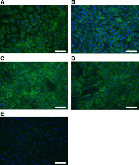
Fig. 1.

Cytokeratin immunostaining of cultured BOEC in different number of cell culture passages. Immunostaining with anti-cytokeratin antibody of cultured BOEC in: a passage 0; b passage 1; c passage 2; d passage 3; and e negative control. Goat anti-mouse-IgG DyLight 488 conjugate (green) was used for staining cytokeratin as a secondary antibody. DAPI (blue) was used to visualize nuclei. Magnification was set at 200 X. Bar in each figure represents 100 μm. Representative pictures of BOEC cultured in LOW glucose medium are shown
