Fig. 3.

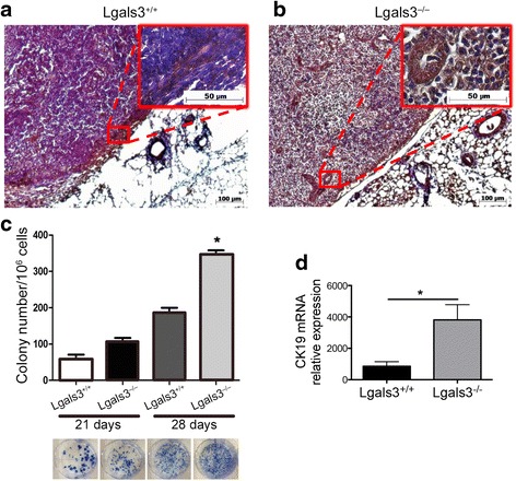
The detection of 4T1-derived metastatic colonies in the lymph nodes is increased in Lgals3−/− mice. Representative immunohistochemical staining of CK-19 in the draining lymph nodes of Balb/c a Lgals3+/+ or b Lgals3−/− mice previously inoculated with 105 4T1 mammary carcinoma cells in the fourth mammary fat pad for 28 days. c Number and representative images of clonogenic 4T1 metastatic cells cultured from a total of 105 draining lymph nodes cells 21 and 28 days p.o.i. d CK-19 mRNA levels in draining lymph nodes cells of Balb/c Lgals3+/+ or Lgals3−/− mice 15 days p.o.i. with 4T1 mammary carcinoma cells. Data are the mean ± S.D., n = 4, three animals per group; *p < 0.05
