Figure 3.
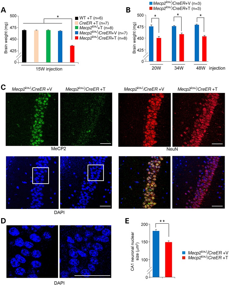
MeCP2 depletion at advanced adult stages leads to rapid shrinkage of the brain and reduction in nuclear size of hippocampal neurons. (A) The brains of the symptomatic 15 W-Tam-injected Mecp2loxJ/CreER male mice are smaller than those of their age-matched control littermates. The brain weights were measured 5–15 days post-injection, when the Tam-injected Mecp2loxJ/CreER mice were severely symptomatic. One-way ANOVA followed by appropriate post hoc for multiple-comparisons test was used to determine differences between groups. Error bars are mean ± SEM. *P < 0.05. (B) The brains of the symptomatic 20 W-, 34 W- and 48 W-Tam-injected Mecp2loxJ/CreER mice become smaller than the brains of the control Veh-injected Mecp2loxJ/CreER mice within days after Tam injection. The Mann–Whitney U-test was used to compare between Veh- and Tam-injected animals of each age group. Error bars are mean ± SEM. *P < 0.05. (C) Representative images of immunostaining of the CA1 area of the hippocampus of 15 W-Tam- or Veh-injected Mecp2loxJ/CreER male mice at their symptomatic stage (score of 6–7, see Fig.1) immunolabeled for MeCP2 (green) and NeuN (red). DAPI (blue) represents nuclear staining. (D) Enlargement of the CA1 area (framed areas in C) showing smaller nuclei in CA1 hippocampal neurons of symptomatic 15 W-Tam-injected Mecp2loxJ/CreER male mice compared to the Veh-injected littermate control male mice. (E) Nuclear size quantification data revealing smaller nuclei of the CA1 hippocampal neurons of the 15 W-Tam-injected Mecp2loxJ/CreER mice at their symptomatic stage (score of 6–7) than those of the 15 W-Veh-injected Mecp2loxJ/CreER control littermate mice. t-test was used to compare within Veh- and Tam-injected groups. Error bars are mean ± SEM. **P < 0.01. n = 3 mice per group. n = 20 neurons per animal. Scale bars, 50 µm.

