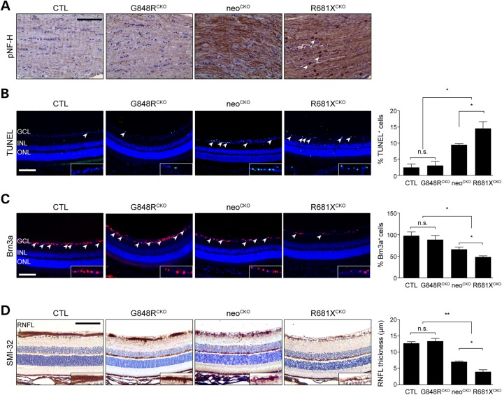
Figure 3.
The germline Nf1 gene mutation dictates the degree of optic glioma-induced retinal dysfunction. (A) pNF-H immunostaining in the optic nerves of control, G848RCKO, neoCKO and R681XCKO mice revealed increased immunoreactivity in neoCKO and R681XCKO mice, with R681XCKO mice exhibiting additional punctate staining (arrowheads). (B) The retinae of R681XCKO and neoCKO mice had increased percentages of TUNEL+ cells (5.9- and 4.8-fold increase, respectively) compared with control mice (n = 5 mice per genotype). (C) The retinae of R681XCKO and neoCKO mice had lower percentages of Brn3a+ cells (50 and 41% decrease, respectively) compared with control mice (n = 5 mice per genotype). (D) The retinae of R681XCKO and neoCKO mice had RNFL thinning as revealed by SMI-32 immunostaining (3.4-and 3.2-fold decrease, respectively) compared with control mice (n = 5 mice per genotype). GCL, ganglion cell layer; INL, inner nuclear layer; ONL, outer nuclear layer; RNFL, retinal nerve fiber layer. Scale bars: 100 µm. All data are represented as means ± s.e.m. (**P < 0.01; *P < 0.05; one-way ANOVA with Bonferroni post-test). n.s., not significant.

