
Figure 4.
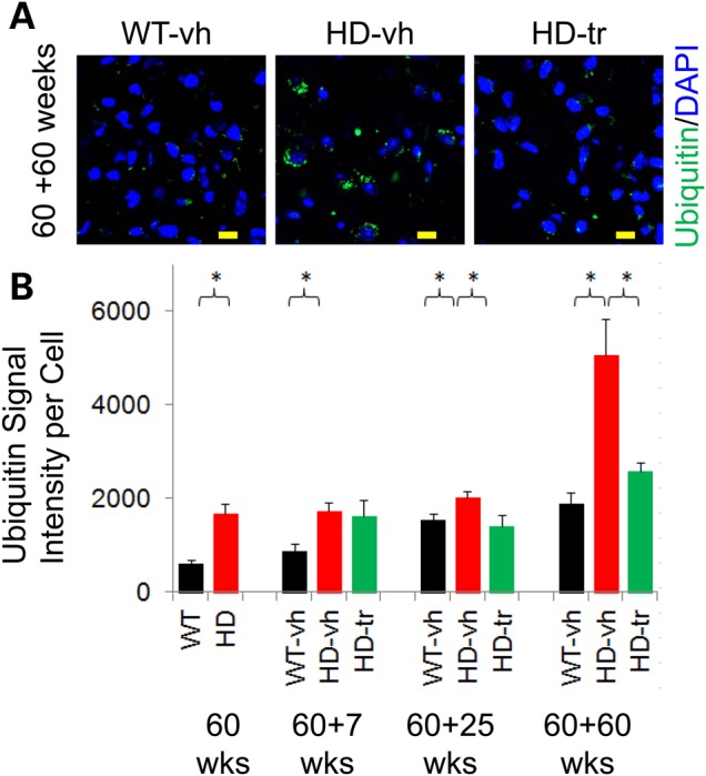
XJB-5-131 treatment of HdhQ(150/150) animals reduces inclusions in the brain. (A) The signal staining intensity for anti-ubiquitin antibody (green) after 60 weeks of XJB-5-131 treatment (60 + 60 weeks). Inclusions are elevated in the striatal cells of HD-vh mice relative to striatal cells WT-vh, and are suppressed in HD-tr by XJB-5-131 treatment. Scale bars indicate 10 µm. Blue is DAPI staining of DNA in the nucleus. (B) Quantified results of (A) for WT-vh (black) HD-vh (red), and HD-tr (green), as indicated. The start of treatment is 60 weeks; the time of treatment is n, as in (60 + n). There are significantly fewer inclusion bodies (measured by IF signal of ubiquitin) after XJB-5-131 treatment in the striatal cells of HD-tr relative to WT-vh and HD-vh and at all measured time points (n = 3 animals per genotype, >250 cells each) (*P < 0.05, Student's t-test, one-tailed homoscedastic).